| Shell |
|
Overview 3D anatomy Digestion Senses Nerves Respiration Circulation Reproduction Other Shell |
The shape of the shell in genus Pomacea:
The shell of most apple snail species is oval, subglobosely or globosely
conic, while some species (like Marisa cornuarietis) have a rather
discoidal shell (flat shell). Apple snails have a dextral shell
(shell opening at the right like in the picture above) except the apple snails
from the genus Lanistes who have a sinistral shell (shell
opening at the left), although there are reports of sinistral animals in the
other genera (see the literature: CAZZANIGA,
N. J. & ESTEBENET, A. L. 1990) and recently the author
of this website discovered a sinistral Marisa cornuarietis
snail in his aquarium. Another sinsitral shell (Pila ampullacea)
is in the Dr. Harry G. Lee collection.
The Lanistes snail has a sinistral shell, but the body is dextral. This phenomenon is known as "hyperstrophic" sinistral. In this case the shell grows upward instead of downward, making the umbilicus inverted to the outside. In a truly sinistral snail, the shell and the body are turning to the left and in such cases all organs are inverted (for example the lung would be at the right instead of at the left body side). In a hyperstrophic sinistral snail, the body of the snail dextral. This dextral body becomes clearly visible during mating. Especially of you compare this with a dextral snail like Pomacea diffusa: check out the Lanistes lybicus page for pictures. The evolution of a dextral to a hyperstrophic sinistral snail is showed in the movie below (at the right). Halfway the inversion from dextral to hyperstrophic sinistral one can recognise the planorbid shape of the Marisa genus.
|
|
 Dextral to hyperstrophic sinistral evolution. |
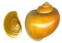
A whorl (coil) is a complete turn of the shell. The shape of the whorls
varies from oval in Afropomus, Lanistes,
Marisa, Pila,
Pomacea, and Saulea to neritoid (drop-shaped) in Asolene
and Felipponea and increase fast in diameter from
the shell top to the shell opening. The last whorl or body whorl is rather big
due to the space needed to encompass the lung. The sutures are the lines
on the shell surface where two adjoining whorls meet. The sutures differ from
species to species: some have a straight 90� angle (like in Pomacea
diffusa) others have intended sutures (like in Pomacea
canaliculata) or have nearly flat sutures (more than 90� angle between
the whorls) (like in Pomacea flagellata flagellata).
The umbilicus (the deep pit in the centre of the shell) can vary from
closed (imperforated), like in the Afropomus
balanoideus, to rather wide (perforated), like in the Marisa
genus.
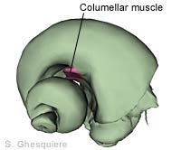
The columella is the central axis of the shell (from shell-top to umbilicus).
The snail's body is attached to shell with the columellar muscle, which
is attached to the columella. The columellar muscle is also known as the retractor
muscle, as it enables the snail to retract firmly into the shell. If the columellar
muscle breaks, the snail loses its shell and dies.
|
|
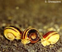
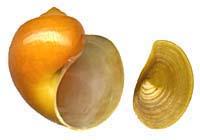
The shell colour varies from white, yellow-brown, brown to greenish-brown,
with or without reddish or brownish spiral surface bands. The pigments that
determine the shell's colour are situated in the organic layer or periostracum at the shell surface.
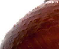
The surface of the shell can be smooth or rough, depending of the species
and the environment.
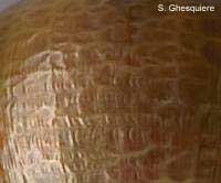
Malleation (hammered surface) occurs mainly
when the snail grows fast. In such cases the anorganic periostracum, which is
first created, is very thin and wrinkles before it becomes enforced with the
rigid ostracum and hipostracum.
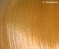
Stria (growth lines) are formed at slower growth.
New shell material is deposited bit by bit at the shell opening, creating the
a new small ridge or stria with every new piece added.
|
|
|
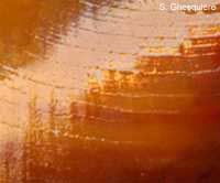
On the shell of young snails and in the case of some species on the adult shells as well, there are spiral rows of small hairs (periostracal hairs). These can be seen in reflecting light on a wet shell.
|
|
|
The shell lip (the margin of the shell at the shell opening) is often
thickened in mature snails, while it is sharp in younger snails. The lip is
curved at the outside, while rather straight near the umbilicus.
The operculum (the shell door) is a corneous
plate (except in the genus Pila, where the operculum
is calcified at the inside during the life of the snail), with a concentric
structure and a nucleus near the parietal margin (close to the umbilicus). With
this shell door, the snail is able to close of its shell to survive periods
of drought and as protection against predators.
|
|
|
|
|
The structure of the shell:
The shell consists of several layers. The layer on the outside is an organic
layer, build up of several layers conchioline, which contains the pigments which
make up the colour of the shell. This layer, also called the periostracum,
isn't as strong as the underlying layer, the prismatic layer. The prismatic
layer or ostracum is a strong, calcium rich layer that normally doesn't
contain pigments and has a white colour. However, the pigments from the periostracum
can slowly migrate to the prismatic layer. On older shells, the prismatic layer
becomes sometimes visible because the periostracum tends to get little holes
and is sometimes almost completely lost at the top of the shell. A picture of
shell with little holes in the periostracum can be seen at the 'Snail
diseases' section.
The calcium carbonate crystals of this layer are oriented perpendicular to the
shell surface. The smooth layer at the inside of the shell is the pearl layer
or hipostracum. The calcium carbonate crystals of the hipostracum are
oriented parallel to the shell surface. The pearl layer is attached to the prismatic
layer with a very thin layer of conchioline. The thickness of the shell depends
on the species, the age of the snails and is also influenced by environmental
conditions like the calcium concentration of the water.
 Shell of Pomacea diffusa, view of the inside to illustrate the shell construction. |
 Computer animation of a growing shell. |
When a snail grows, the shell has to be enlarged to fit the snail's body.
To accomplish that, a snail gradually extents its shell by adding new parts at the shell
opening.
The growth process is carried out in two stages: In the first stage the thin,
transparant and organic outer layer (periostracum) is created, on which the
calcified inner layers are deposited in the second stage. The shell material
is secreted by specialized cells of the mantle.
Apple snails can grow their shell very fast under the right conditions: peak
rates of 0.5 cm (0.2 inch) new shell a day do occur.
The computer animation above illustrated the growth of a shell. The oldest part of the
shell is located at the top, while the most recent part is located near the shell opening
(aperture).
When a new shell part is added, small vertical lines (transverse
stria) are formed on the surface of the shell.
The thickness and regularity of those lines varies with the environmental conditions,
the age of the snail and the species. They
provide information about the environmental conditions during the snail's life
(similar to what the grains of wood tell us about the life of a tree).
The operculum or trapdoor of apple snails grows in a similar way as the shell:
a new material is added in a circular fashion. The result is a concentric operculum
with the oldest part in the center. The operculum is only enforced with a calcium
layer in the genus Pila, the other genera have
a horny operculum.
Due to the way snails solve the growth problem (enlarging their shell), they always have
to carry the whole construction with them, even if they do not inhabit the oldest whorls.
Crustaceans and insects, for example, use another option to overcome the growth problem:
they regulary shed their whole skin when they grow and replace it with a larger one. The
advantage of that method above the snail-grow method is that they do not have to carry
around obsolete body parts. On the other hand, a crustacean or an insect is a very
vulnerable when it has shed its old skin and the new skin still has to harden.
Calculate your snail's shell volume:
The mathematical formula (the formula of a cone model) used for this calculation / estimation:
|
Volume = 1/3Pi * (Shell width/2)^2 * Shell height |
|
|
|
|
Except where otherwise noted, this page is licensed under a Creative Commons Attribution-NonCommercial-ShareAlike 2.5 License . http://www.applesnail.net |
|