| Embryology: Stage I | |
|
|
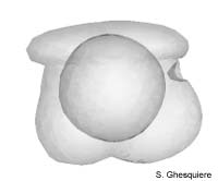 Embryo in Stage 1. |
|
Interactive 3D-models (Java): |
After the gastrulation, stage one starts. This is the first stage out of 12
before the embryo hatches from the egg.
The embryo is about 120µm in case of a Marisa cornuarietis embryo,
while a Pila globosa embryo is somewhat larger (190µm) in size.
The time after deposition of the eggs, at which the first stage starts, mainly
depends on the temperature. At a temperature of 15 to 20°C it takes 3 days
for an embryo of Marisa cornuarietis to enter stage one, while it takes only
28h at a temperature of 25 to 30°C. Between 20 to 25°C (room temperature)
both Pila globosa as well Marisa cornuarietis finish the gastrulation
and start with stage one after 36h.
In this stage, the embryo is bilaterally symmetrical, both internally as well
externally.
On the dorsal side of the embryo the velum is already present. This cap-like
structure is a common feature in the embryonic development of molluscs. In mollusc
that have a larval stage, the velum functions as a swim organ. In apple snails
(Ampullariidae), the velum doesn't have such clear propulsion function, although
the embryo makes circular movements inside the egg at stage 2 and later. The
velum covers nearly half of the embryo at the dorsal side and is marked with
a ciliated band. This ciliated, transverses the whole embryo and consist of
a double row of ciliated cells, also called the prototroch or preoral ciliated
band. At the dorsal side, the velum consists of a ciliated cell plate: the apical
cell plate.
At the ventral-posterior side of the embryo, a patch of cells becomes visible
in the ectoderm. This plate like structure is the anal cell plate.
The pedal cell plate becomes conspicuous at the antero-ventral side. The pedal
cell plate can be considered as a rudimental foot.
The shell plate, a group of cells that will develop into the shell gland in
the future, is present at the posterior side of the embryo, between the velum
and the anal cell plate. At the outside, the shell plate isn't visible yet.
Inside the embryo, the endodermal sac, with its irregular archentric cavity,
is visible. This endodermal tissue has been formed during the gastrulation,
at which parts of the ectoderm are internalised. The posterior side of the archentric
cavity slightly protrudes ventrally to form the future intestines in a later
stage. The whole archenteron has a yellow colour due to the high yolk content
of the cells.
In the mouth region the stomodaeum starts to develop and is visible as a slightly
invaginated cell plate just below the velum and the foot rudiment.
The anus is not yet present at the other end of the alimentary tract in case
of Marisa cornuarietis, while it has been reported to exist in this stage
in Pila globosa (Ranjah, 1942).
|
|
|
|
Except where otherwise noted, this page is licensed under a Creative Commons Attribution-NonCommercial-ShareAlike 2.5 License . http://www.applesnail.net |
|