| Embryology: Stage IV | |
|
|
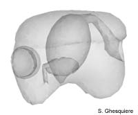 Embryo in Stage 4. |
|
Interactive 3D-models (Java): |
During previous stage, the Marisa embryo has grown steadily, resulting
in a size of 240µm at the start of this stage. The the Pila globosa
embryo is, however, only sligthly enlarged compared to stage III and only measures
240µm in length at stage IV. Appearantly the Marisa embryo has
catched up in terms of growth.
Whereas the embryo was still symmetrical externally in the previous stages,
this stage is marked by the lost of this external symmetry because the shell
rudiment has shifted to the left side of the body. The border around this shell
plate (the rudimental shell gland) has thickened and will develop into the future
mantle edge.
The stomodaeum has become more conspicuous, in particular at the front side,
where the mouth region is differentiated, with a buccal cavity at the front
side and a narrowing at the back side, forming the oesophagus.
Inside the buccal cavity, the radular sac starts to develop, although it's only
visible as a invagination on the floor of the buccal region. The posterior part
of the stomodaeum, which communicates with the primitive stomach, has shefted
to the right, contributing to a further asymmetry of the embryo.
Albuminous fluid (yolk) can be found inside the archenteron or primitive stomach
in this stage. This is not suprisingly as the stomodaeum provides an opening
for the intestinal system. Nevertheless, it illustrates that the stomodaeum
and oesophagus already function in the sense that they are capable to transport
fluid. There are, however, no muscular movements visible, which suggest that
the transport of fluid is driven by the cilated current on surfaces of the intestinal
system.
The endodermal sac, which encompasses the primitive stomach, is becoming pear-shaped
in this stage, with the narrowing part directed at the ventral-posterior side.
The pericardium, heart and kidney still consist of one common rudiment, but
become more divided due to constrictions of the wall. At the end of stage IV,
both the pericard and the kidney are distinguishable, although still closely
connected. The kidney itself now communicates with the ectodermal invagination
that develops to the renal vestibule, with the ureter in between.
|
|
|
|
Except where otherwise noted, this page is licensed under a Creative Commons Attribution-NonCommercial-ShareAlike 2.5 License . http://www.applesnail.net |
|