Embryology: Stage II
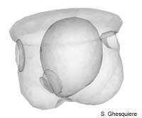
 Back

OverviewEggEarly cleavagesStage 1Stage 2Stage 3Stage 4Stage 5
Stage 6Stage 7Stage 8Stage 9Stage 10Stage 11Stage 12


Embryo stage 2
Embryo in Stage 2.


3d-models

Interactive 3D-models (Java):
- Model of embryo in stage 2


After about 40h, stage II of the embryonic development starts. In this stage the Marisa cornuarietis embryo is about 160µm in length, while the Pila globosa embryo measures 210µm.
While the embryo did not move much in the previous stage, it now makes circular movements inside the egg. These movements are mainly caused by the ciliar waterflow on the velum.
At the front side of the embryo, the foot rudiment or pedal cell plate has become more prominent as the cells in this region have increased considerably both in numbers as well in size.
The anal cell plate at the posterio-ventral side of the embryo is enlarged as well. The shell plate or rudimental shell gland is visible above the anal cell plate as a slightly invaginated, thickened plate. The area around the shell plate and the anal cell plate will develop in the future visceral sac that will ultimately become embedded in the adult shell.
Besides the shell plate, another cell plate appears: the rudiment of the ureter and the renal vestibule. While the shell plate is well visible, this cell plate is small and rather difficult to observe.
On the dorsal side of the velum, two new cell plates develop anterior of the apical cell plate: the head plates. These thickened cell plates, one on each side, are easy to distinguish due to the heavily granulated cells of these plates. During the embryonic development, these head plates will differentiated and develop into the cerebral ganglia, the tentacles and the eyes.
The stomodaeum or rudimental mouth further invaginates during stage II, but does not make contact with the primitive (or larval) stomach or archenteron.
The archenteron, the cavity inside the embryo, surrounded with endoderm, enlarges in this stage. Near the anal cell plate at the posterior side of the embryo, the rudimental intestine grows slowly as protrusion of the archenteron.
One of the more remarkable events in this stage is the formation of two mesoderm plates that become conspicuous at the posterior side of the embryo, at each side of the endodermal sac (the archenteron/larval or primitive stomach). These cell masses that form the endoderm are unequally in size, the left being smaller then the right one. The right mesodermal plate will differentiate further in the next stages to form the heart and the kidney, while the left one disappears during development.
The origin of the mesoderm in apple snails (Ampullariidae) appears to be teloblastic.


Top
Top


Creative Commons License
Except where otherwise noted, this page is licensed under a
Creative Commons Attribution-NonCommercial-ShareAlike 2.5 License
.

http://www.applesnail.net