
|
|
|
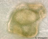
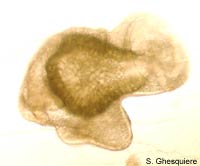
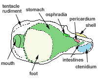
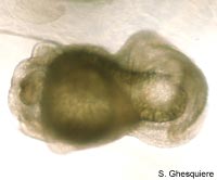
 Marisa cornuarietis embryo at day 12. At the right is the cup-like shell, at the left bottom the foot by which the snail walks inside the egg. The tentacles are still under-developed, and the eyes aren't visible yet. |
The embryology section of this website is currently under development. As one
can see at the menu structure above, there is quite a lot to tell about the
embryology of apple snails.
However, it will take some time to write the text, make illustration and pictures...
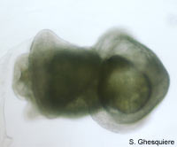
The embryotic development of apple snails. In this case of Marisa cornuarietis as the embryos in this species are well visible inside the eggs and therefor this snail is a very easy animal to study the embryotic development process.
Note that this page is still under heavy development as well.. and it's far from complete and probably contains quite a lot of errors... :-)
Day 0
The eggs are deposited on a surface or object in the water. The eggs come out
of the snail's body, one by one in a long string that folds up in such way that
the eggs are attached to each other in one clutch.
At this stage the eggs are small and have a white, translucent appearance.
 Marisa cornuarietis depositing the eggs. |
Day 2
The eggs slowly enlarge when they take up more and more water and they are
more and more transparent.
Inside the eggs, the little embryos become visible as very small white spots
on the walls of the eggs.
 Eggs in a gelatinous mass, 2 days old. |
Day 4
The little embryos can now be seen as small white spots moving around inside
the eggs.
With the naked eye, it's impossible to see more than a spot, but under a microscope
the development process is clearly revealed to the eye.
One of the first things that attracts attention is the circular motion these
embryos make and with further magnification one can see a region with cilia
that creates a water flow that propels the snail around in the fluid.
The large oval cavity at the center of the embryo is also a striking feature
in this stage of development. The shell is only
visible as a very small round depression at the top of the embryo. The foot
is no more then a bulge and no eyes or
tentacles are visible.
At the bottom-front of the embryo one can see a large mouth
opening. The rectum is located somewhere
at the back, as well are the developing intestines.
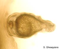
 Marisa cornuarietis embryo inside the egg at day 4. The size of the embryo is about 350µm in diameter. |
||
 Close up of the embryo, left side view. The different areas of the snail body are already forming in this stage. |
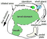 A ciliated area produces a water flow that propels the embryo inside the egg in a circular motion. |
 -- |
 Front view of the same embryo. The large mouth opening in the center. Note that this photograph is upside down. |
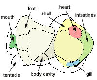
Day 7
The shell moves to the left side of the snail
body and is enlarged and visible as a round plate with an elevated border around
it. This border around the shell plate is the growth border that enlarges the
shell by adding new material.
The intestines as well as the
mouth region are more and more differentiated and illustrated by the increased
details in these organs. The foot
also grows and shows some small movements whenever the snail bumps on the walls
of the egg. However, the swimming with the ciliated area on the head is still
the most important method of moving around for the embryo.
One of the most interesting processes in this stage are the first visible heartbeats.
The heart itself is not very well visible,
but one can see slow contractions at the end of the body cavity. Once every
3-6 seconds a slow contraction takes place.
 Left side view. At this stage, the embryo gets a more distinguished foot and shell. The size of the embryo is about 400µm. |
 The shell has moved from the top of the body to the left side and is now visible as a cup-like plate at the tail. |
 -- |
 Bottom view of the embryo. The shell plate is visible at the side of the snail's body. |
 The first heartbeats become visible through the microscope. |
 -- |
Day 9
At day 9 the eggs are completely transparent and are about 5 mm in diameter.
The small snails (450µm length) are visible with their cup-like shell
on their back.
The shell of the embryo has now grown from a plate to a cup-like structure that
engulfs the posterior part of the snail body.
Heartbeats are clearly visible and the
heart now consists of two parts (atrium and ventricle) and is closely connected
with the end of the central body cavity at the shell side. The walls of the
central cavity are very thin at this place, whereas they are much thicker near
the snail head.
The position of the intestines
becomes more and more visible while the rectum is moved toward its final place
near the shell border at the right side of the
snail.
Eyes are not yet developed, but the tentacles
are already there in the form of small bulbs at the side of the head, which
in turn is also more differentiated, as well as the mouth region. In the latter,
the rasp tongue or radula is already existing although rather rudimental.
With the foot better developed, the
snail can move over the inner surface of egg instead of the swimming movement,
propelled by small currents created by cilia on the head. Nonetheless, the cilia
movements are still there, more exactly on the head and the shell border. In
later life, these water currents become important to create a constant flow
through the mantle cavity,
from the siphon at the left side
over the gills towards the right
side of the body.
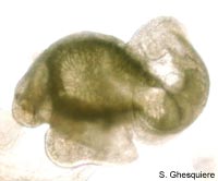 Left side view. Size of the embryo: 450µm length |
 The shell is now a cup-like structure. |
 -- |
 Bottom view of the embryo. |
 The tentacles are developing. |
|
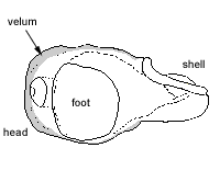
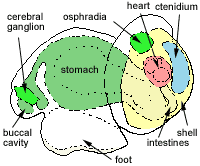
Day 11
The posterior (tail side) part of the central body cavity is enlarged compared
to the situation in the days before. The thinning in the posterior wall at the
side of the heart is smaller, but still
visible. The cavity also starts to coil a bit to the left at shell
side (see the underside view below).
Due to the further differentiation of the organs and hence the more detailed
anatomy, it becomes more difficult at this stage to distinguish the different
structures.
An example of such hard to see and comprehend organ is the developing kidney
above the now well visible heart and the development of the stomach.
The shell now envelops a large part of the body and the shell door (operculum)
is visible as a small plate on top of the foot.
A structure what once will become a gill
starts to develop and is somewhat visible at the roof of the shell.
The tentacles are still small and
the eyestalk is still absent, although
a small bulb near the tentacles becomes visible. The eyes themselves are not
present at this stage.
 Left side view. Note the more and more detailed structure of the organs. Size of the embryo: 500µm length. |
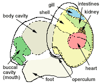 The shell door (operculum) starts to develop. |
 Bottom view of the embryo. Interesting to see is the start of the coiling process as the posterior part of the body cavity starts to bend. |
 The tentacles are there, but the eyes are still absent. |
|
|
|
|
Except where otherwise noted, this page is licensed under a Creative Commons Attribution-NonCommercial-ShareAlike 2.5 License . http://www.applesnail.net |
|